• Skip to main content
  • Skip to footer

Dr Dharambir S Sethi

24 by 7 Appointment
+65 8125 3580

MAKE AN APPOINTMENT
MAKE AN ENQUIRY
  • HOME
  • ABOUT A/PROF SETHI
    • About A/Prof Sethi
    • International Meetings
    • Photo Gallery
  • MEDICAL CONDITIONS & PROCEDURES
        • Allergy Services
          • Allergic Rhinitis
        • Nose & Sinuses
          • Running Nose
          • Nose Bleeds
          • Nasal Polyps
          • Sinusitis
          • Why do I need a CT scan for my sinuses?
          • Eustachian Tube Dysfunction (ETD)
          • Nose Cancer
          • Nasal Septum
          • Nasal Turbinates
          • Nasal Endoscopy
          • Radio-frequency Reduction
          • Endoscopic Sinus Surgery
          • Antral Wash Out
        • Ear Diseases & Hearing Loss
          • How Do We Hear Sound
          • Otoendoscopy
          • Ear Pain
          • Ear Wax
          • Keratosis Obuternas
          • Otomycosis
          • Acute Otitis Media
          • Bullous Myringitis
          • Glue Ear
          • Acute Ear Pain
          • Cholesteatoma
          • Pulsatile tinnitus
        • Neck, Submandibular Thyroid & Parotid Gland
          • Lymph Nodes
          • Parotid Tumours
          • Submandibular Masses
        • Throat and Voice
          • Adenoids
          • Gastro-oesophageal Reflux Disease
          • Tonsils
        • Snoring and Sleep Apnoea
          • Sleep, Snoring and Sleep Apnoea
        • ENT Problems in Children
          • Nasal & Septal Surgery
        • Minor Endoscopic Procedures
        • Advanced Endoscopic Sinus Surgery Procedures
          • Functional Endoscopic Sinus Surgery
          • Cerebrospinal Fistula
          • Optic Nerve Decompression
          • Endoscopic Pituitary Surgery
          • Endoscopic Repair of Encephalocele
        • Endoscopic Skull Base Surgery
  • PATIENT INFORMATION
    • Medical concierge
    • ENT Emergencies
  • BLOG
  • CONTACT
    • Make an Appointment
    • Make an Enquiry
    • Getting to Our Clinic

Home / Nose Sinuses / Why do I need a CT scan for my sinuses?

Why do I need a CT scan for my sinuses?

Call +65 8125 3580
for 24 by 7 appointment

MAKE AN APPOINTMENT
MAKE AN ENQUIRY

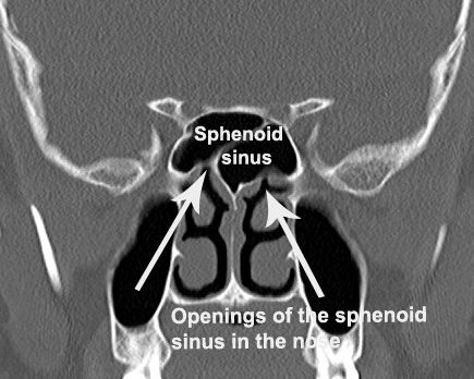
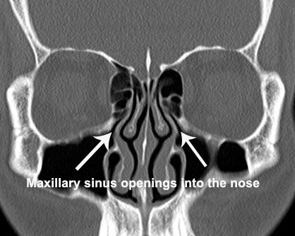
  • The Maxillary Drainage into the nose
  • The Sphenoid Sinus Drainage
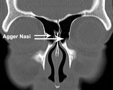
  • The Agger Nasi
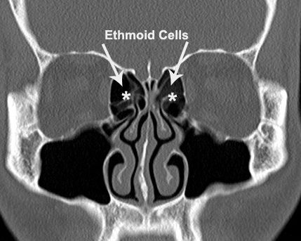
  • The Ethmoid Sinuses
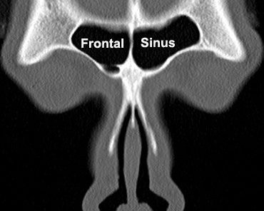
  • The Frontal Sinus
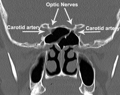
  • The Optic Nerves and the Internal Carotid Artery
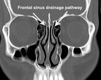
  • The Frontal Drainage Pathway

If you have recently consulted a Ear Nose & Throat doctor for nose and sinus related symptoms and the doctor has suggested a CT scan of your sinuses here is the reason why. Computed tomography, more commonly known as a CT or CAT scan, is a diagnostic test that produces multiple images or pictures of the inside of the body. The cross-sectional images generated during a CT scan can be reformatted in multiple planes, and can even generate three-dimensional images. Unlike the traditional X-rays which provide very little information, CT scans are preferred because of the detailed information the CT images provide. CT scans is the imaging modality of choice for evaluating the paranasal sinuses. CT of the sinuses is primarily used to detect the presence of disease which may be inflammatory, infective or a growth within the sinuses. The paranasal sinuses are hollow, air-filled spaces located within the bones of the face and surrounding the nasal cavity. These sinuses connect with the nasal cavity trough very narrow channels a system of air channels connecting the nose with the back of the throat.

Fig-01
Figure 1

There are four pairs of sinuses, each connected to the nasal cavity by small openings. The sinus in the forehead is the “frontal sinus” shown as FS in the Figure 1, the sinus in the cheek bone is called the “maxillary sinus” shown as MS, and multiple small sinus between the eyes and the nose are called the “ethmoid sinuses” shown as ES and the sinus located deep in the skull behind the nose and somewhere in the middle of the skull is the “Sphenoid Sinus” shown as Ss in the figure 1. On the CT scan “air” appears black and the “bone” white on the images. This provides an excellent contrast which enables detection of any disease with in the sinuses. The white arrow shows the contents of the right eye are proplapsing into the right ethmoid sinus. The information is critical to the surgeon so that he can take precautionary measure to avoid a major complication to the eye. Similarly in  figure 4B the white arrow is pointing to the optic nerve which is the nerve of vision. Notice that the nerve is literally floating in the sinus. Without the CT scan the surgeon would be completely oblivious to this situation. Damage to nerve may render the patient blind.   CT scans also provide information that enables us to differentiate whether the sinuses are full of inflammatory disease, fungus, fluid, mucoid secretions or tumors. Sometimes contrast material may be used for this purpose .

Fig-02
Figure 2.

In the event there is any inflammation of the mucosal lining, the mucosa becomes swollen and will appear as thickened “grey” lining as shown in figure 2A (white arrow). If the disease is quite extensive the entire sinus becomes “grey”. (Figure 2B: Note the left maxillary sinus is completely greyed out. This may be due to inflammation, infection, pus, fungus or evena growth with in the sinus.

Fig-03
Figure 3

As just one, few or all the sinuses may be involved with the disease process, CT scans of the sinuses will also provide information on the extent of the disease. This information is crucial in planning the surgery.In figure 3 all the sinuses are involved in the disease process.Figure 4

F3
Figure 4

The CT scan is also akin to a “road map.” It provides information of the pertinent anatomy of the sinuses. Anatomy of the sinuses is not fixed. It varies from person to person. In fact, it can be vary on different sides of the same person. This information is important to avoid some critical structures such as the orbit, brain, optic nerves and major blood vessels during the surgery.In figure 4A the contents of the right eye are prolapsing into the ethmoid sinuses because of a “dehiscence” of a bony defect in the partition between the two. This condition if not recognised before the surgery predisposes the eye contents to injury during the surgery. The injury can be serious enough to result in blindness. In figure 4B the arrow points to the right optic nerve which is literally “floating” in the right side of the sphenoid sinus. Again if not recognosed before the surgery the nerve could be damaged during the surgery resulting in blindness.

I am sure by now you will understand of the importance of CT scan particularly when surgery is contemplated. It is to “get to know your sinuses better!!”

NOSE & SINUSES CONDITIONS

  • RUNNING NOSE
  • NOSE BLEEDS
  • NASAL POLYPS
  • SINUSTITIS
  • NOSE CANCER
  • EUSTACHIAN TUBE DYSFUNCTION (ETD)
  • NASAL SEPTUM
  • NASAL TURBINATES
  • NASAL ENDOSCOPY

NOSE & SINUSES PROCEDURES

  • WHY DO I NEED A CT SCAN FOR MY SINUSES
  • NASAL ENDOSCOPY
  • RADIO-FREQUENCY REDUCTION
  • ENDOSCOPIC SINUS SURGERY
    • Post Operative Care
    • Complications of Endoscopic Sinus Surgery
  • ANTRAL WASH OUT

Footer

MAIN CLINIC

Mount Elizabeth Novena Specialist Centre
#04-21/22/34,
38 Irrawaddy Road
Singapore 329563

  • ABOUT A/PROF SETHI
  • Patient Information
    • Medical Conditions
    • ENT Emergencies
  • MEDICAL CONDITIONS & PROCEDURES
    • Allergy Services
    • Nose & Sinuses
    • Ear Diseases & Hearing Loss
    • Neck, Submandibular Thyroid & Parotid Gland
    • Throat and Voice
    • Snoring and Sleep Apnoea
    • ENT Problems in Children
    • Ear Nose and Throat Emergencies
    • Minor Endoscopic Procedures
    • Functional Endoscopic Sinus Surgery
    • Advanced Endoscopic Sinus Surgery Procedures
    • Endoscopic Skull Base Surgery
    • Nasal & Septal Surgery
  • CONTACT US
    • Make an appointment
    • Make an enquiry
    • Getting to our clinic

Sitemap • Privacy Policy

© Copyright 2020 Dr Sethi. All Rights Reserved.