Home / Neck, Submandibular Thyroid & Parotid Gland / Submandibular Masses
Submandibular Masses
Call +65 8125 3580
for 24 by 7 appointment
What are swelling below the jaw?

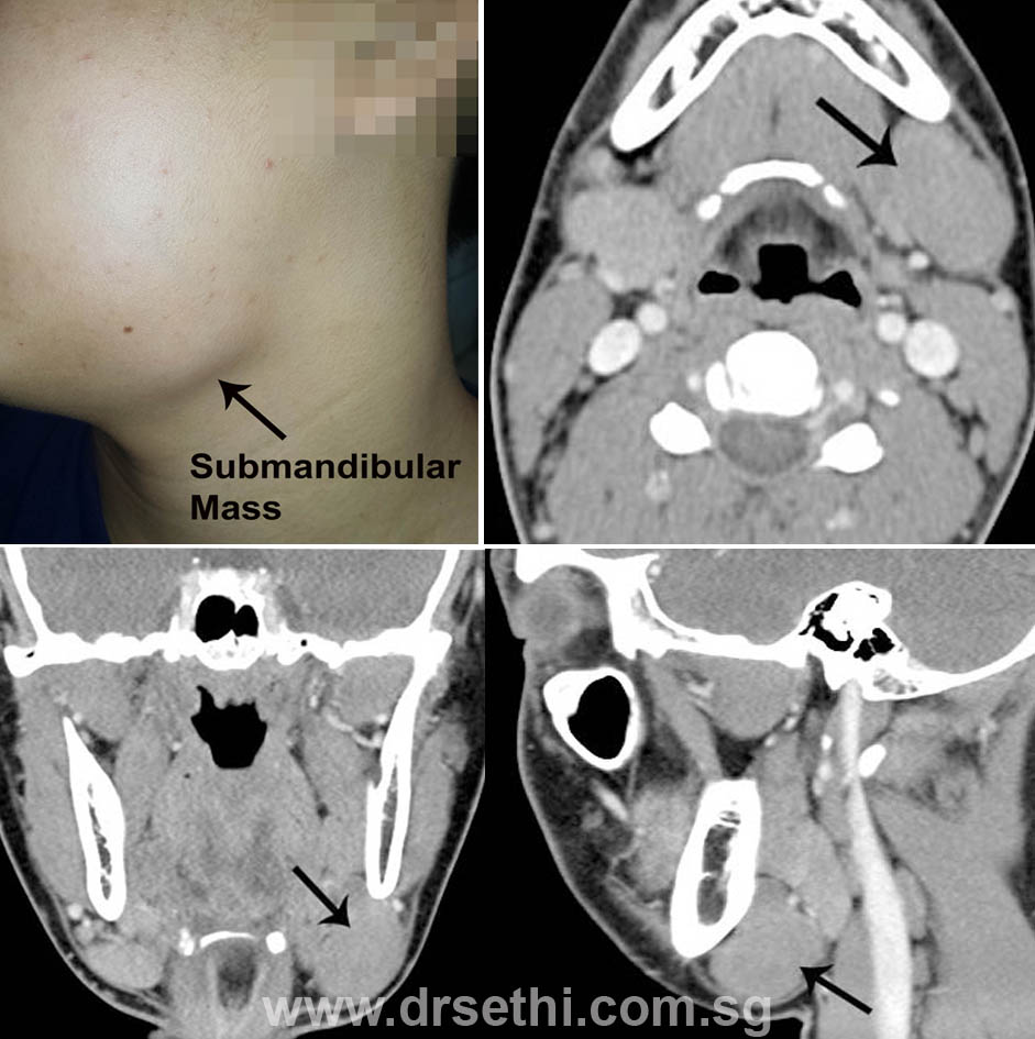
Located just below the mandible (jaw) and extending from the angle of the mandible to the midline is a triangular area that contains the submandibular gland. The submandibular gland is the second largest of the salivary glands next to the parotid gland. Most of the swelling or masses, in this area are related to the submandibular gland. These may be infective in nature or may be tumours. The submandibular triangle also contains several lymph nodes. An enlarged lymph node in this triangle may sometimes be mistaken for an enlarged submandibular gland.
Any swelling in this region needs to be investigated. A detailed history may provide clues as to whether the swelling is infective or may be a growth. It will also provide information regarding the possible pathology.
Examination will include examination of the neck to rule out other swellings and to determine the possible extent and consistency of the swelling. Often the doctor may do a bimanual examination which involves feeling the swelling both from inside the mouth and the neck. Endoscopic examination of the nose and throat is an essential part of the clinical examination.
Imaging studies such as CT scan of the neck or MRI of the neck will provide information regarding the extent and relationship of the swelling with the structures around it. This information is critical in planning surgical excision should it be necessary.
A fine needle aspiration (FNAC) which involves aspirating some cells from the swelling to have a histological diagnosis may be helpful.
How is a submandibular gland excised?

If you have been diagnosed to have a SUBMANDIBULAR GLAND TUMOR based on your clinical examination and other investigations, you will need to have it excised.
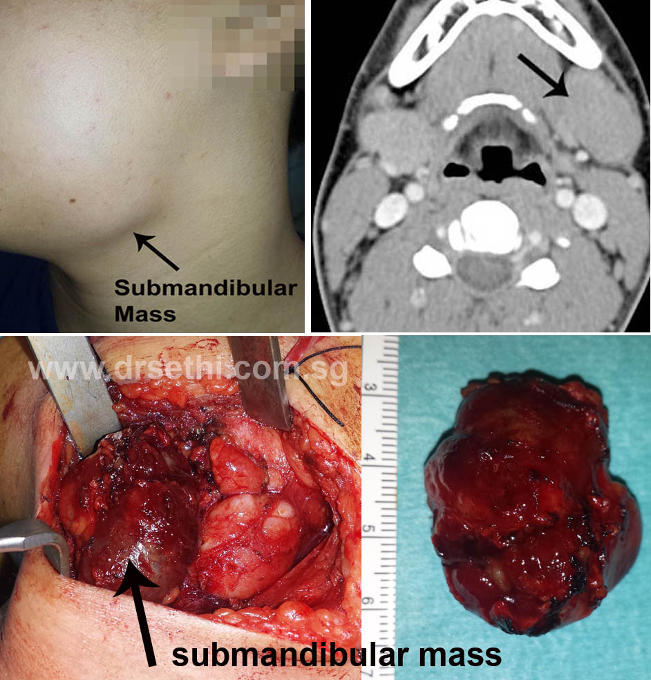
Most of the tumors in the submandibular gland are benign but have to be excised for a definitive diagnosis. Furthermore some of these tumors may be potentially malignant necessitating surgery. The surgery involves excision of the submandibular gland.
The surgery is performed under general anesthesia.
An incision is made along the upper neck crease about 2 cm below the jaw. The incision usually heals very well and the scar is barely perceptible after a few months.
A nerve runs in this region is the marginal mandibular branch of the facial nerve that supplies that part of the angle of the mouth. Though great precaution is taken to protect this nerve, sometimes trauma to the nerve as a result of instrumentation may result in temporary or permanent weakness of the angle of the mouth. The submandibaular gland or the mass is carefully dissected from the surrounding structures which include nerves, veins, arteries and msucles. The surgery may take close an hour or an hour and half.
A small tube is usually left in the wound for 2-3 days to drain any blood clots and prevent a hemotoma. The incision may be closed using subcuticular sutures or interrupted skin sutures depending on the preference of the surgeon.
What to expect after the surgery:
You may experience some pain and discomfort after the operation. These symptoms usually resolve after about 24 hours’
You may have to stay in the hospital for 2-3 days after the operation. This depends of the complexity of the surgery and the drainage from the wound. Usually by second or third the drainage is minimal and the drain can be removed.
Your first follow up appointment is usually in a week to remove the stitches.
The histology of the excised mass is usually available within 3-5 days.
