• Skip to main content
  • Skip to footer

Dr Dharambir S Sethi

24 by 7 Appointment
+65 8125 3580

MAKE AN APPOINTMENT
MAKE AN ENQUIRY
  • HOME
  • ABOUT A/PROF SETHI
    • About A/Prof Sethi
    • International Meetings
    • Photo Gallery
  • MEDICAL CONDITIONS & PROCEDURES
        • Allergy Services
          • Allergic Rhinitis
        • Nose & Sinuses
          • Running Nose
          • Nose Bleeds
          • Nasal Polyps
          • Sinusitis
          • Why do I need a CT scan for my sinuses?
          • Eustachian Tube Dysfunction (ETD)
          • Nose Cancer
          • Nasal Septum
          • Nasal Turbinates
          • Nasal Endoscopy
          • Radio-frequency Reduction
          • Endoscopic Sinus Surgery
          • Antral Wash Out
        • Ear Diseases & Hearing Loss
          • How Do We Hear Sound
          • Otoendoscopy
          • Ear Pain
          • Ear Wax
          • Keratosis Obuternas
          • Otomycosis
          • Acute Otitis Media
          • Bullous Myringitis
          • Glue Ear
          • Acute Ear Pain
          • Cholesteatoma
          • Pulsatile tinnitus
        • Neck, Submandibular Thyroid & Parotid Gland
          • Lymph Nodes
          • Parotid Tumours
          • Submandibular Masses
        • Throat and Voice
          • Adenoids
          • Gastro-oesophageal Reflux Disease
          • Tonsils
        • Snoring and Sleep Apnoea
          • Sleep, Snoring and Sleep Apnoea
        • ENT Problems in Children
          • Nasal & Septal Surgery
        • Minor Endoscopic Procedures
        • Advanced Endoscopic Sinus Surgery Procedures
          • Functional Endoscopic Sinus Surgery
          • Cerebrospinal Fistula
          • Optic Nerve Decompression
          • Endoscopic Pituitary Surgery
          • Endoscopic Repair of Encephalocele
        • Endoscopic Skull Base Surgery
  • PATIENT INFORMATION
    • Medical concierge
    • ENT Emergencies
  • BLOG
  • CONTACT
    • Make an Appointment
    • Make an Enquiry
    • Getting to Our Clinic

Home / Advanced Endoscopic Sinus Surgery Procedures / Cerebrospinal Fistula

Cerebrospinal Fistula

Call +65 8125 3580
for 24 by 7 appointment

MAKE AN APPOINTMENT
MAKE AN ENQUIRY

Cerebrospinal fistula: What is a Cerebrospinal Fluid (CSF) Fistula?

Figure 1a:Patient with watery nasal discharge from the left side of the nose

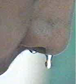
The sinuses have close relationship with the brain. The brain is separated from the nose and the sinuses by thin bone which can sometimes be so thin (or there may be a defect in this bone) that the membranes covering the brain (dura and the arachnoids) may prolapse into the nasal cavity or into the sinuses. These membranes may sometimes rupture causing the fluid that surrounds the brain (cerebrospinal fluid) to leak in the nose. This is called a Cerebrospinal Fluid fistula. Thin watery like nasal discharge especially from one side of the nose and on bending forward may be suspicious of a cerebrospinal fistula (Figure 1a) Such defects need to be closed to prevent the risk of ascending infection from the nose to the brain. The risk of meningitis (infection of the membranes covering of the brain) can be as high as 30%. Diagnosis is usually established from the history, examination of the nasal discharge, and imaging studies. A test called the beta-transferrin is test for the CSF is highly specific and will confirm that the “watery fluid” from the nose is CSF. Imaging studies such as CT and MRI scans will assist in localizing the defect.

Figure 1b: The Ct scan of the patient in figure 1b showing a defect in the skull base
Figure 1b: The CT scan of the patient in figure 1a showing a defect in the skull base (white arrow)


  • CT scan of a patient with a defect in the posterior ethmoid skullbase.
  • 3 year old child with a very large meningoencephalocele. White arrow above shows the defect in the skull base.
  • A very large meningoencephalocele in a 3 year old child presenting as a nasal polyp.
  • CT scan of a patient with a meningoencephalocele. The white arrow above shows the defect in the skull base.
  • MRI scan of the patient with meningoencephalocele
  • CT scan of a patient with CSF fistula with defect in the sphenoid sinus.
  • The CT scan of the patient with CSF fistula showing a defect in the skull base

In the past surgery for closure of CSF defects required an open approach which sometimes included a craniotomy. A craniotomy requires a very large incision on the scalp and removal of the skull bones. The front part of the brain then has to be retracted away to identify the area of the defect. This is then closed with a “pericranial” flap. The surgery though has a good success rate, is also associated with significant morbidity and prolonged hospital stay. In the past 20 years or so endoscopic techniques to repair these skull base defects have been popularised and have an equally good success rate. As the techniques are minimally invasive, there are no incisions made on the scalp or the face, the recovery is faster and the hospital stay shorter. The challenge is to localise the defect pre-operatively using imaging studies such as the CT scan and MRI scan as seen above. Occasionally, the defect may be so small that it may not be identified on the imaging studies. In such cases the use of a dye is useful in identifying the location of the defect. The dye called “fluorescein” is injected in the CSF through a lumbar puncture (intrathecal) before the surgery. Once the defect is identified it can be repaired using tissue taken from the patients own body. This may include fat, cartilage and mucosa. The repair is supported with nasal packing which can be removed in 2 – 3 days. The surgery is done under general anaesthesia and may take about one and half to 2 hours.

Cerebrospinal fistula: How is surgery done for CSF fistula?

https://drsethistagingwpengine.s3-ap-southeast-1.amazonaws.com/Endoscopic+repair+of+encephalocele.mp4

Not sure if your nasal discharge is cerebrospinal fluid fistula? Speak to A/Prof Sethi for a diagnosis at +65 8125 3580. Or book an appointment with A/Prof Sethi for a consultation on your symptoms and treatment options.

ADVANCED ENDOSCOPIC SINUS SURGERY PROCEDURES

  • CEREBROSPINAL FISTULA
  • OPTIC NERVE DECOMPRESSION
  • ENDOSCOPIC PITUITARY SURGERY
  • ENDOSCOPIC REPAIR OF ENCEPHALOCELE

Footer

MAIN CLINIC

Mount Elizabeth Novena Specialist Centre
#04-21/22/34,
38 Irrawaddy Road
Singapore 329563

  • ABOUT A/PROF SETHI
  • Patient Information
    • Medical Conditions
    • ENT Emergencies
  • MEDICAL CONDITIONS & PROCEDURES
    • Allergy Services
    • Nose & Sinuses
    • Ear Diseases & Hearing Loss
    • Neck, Submandibular Thyroid & Parotid Gland
    • Throat and Voice
    • Snoring and Sleep Apnoea
    • ENT Problems in Children
    • Ear Nose and Throat Emergencies
    • Minor Endoscopic Procedures
    • Functional Endoscopic Sinus Surgery
    • Advanced Endoscopic Sinus Surgery Procedures
    • Endoscopic Skull Base Surgery
    • Nasal & Septal Surgery
  • CONTACT US
    • Make an appointment
    • Make an enquiry
    • Getting to our clinic

Sitemap • Privacy Policy

© Copyright 2020 Dr Sethi. All Rights Reserved.